Anatomy Diagram Rib Area : Rib Bone Anatomy Quiz : The rib cage is often simplified as an oval shape.. Each pair is numbered based on their attachment to the sternum, a bony process at the front of the rib cage which serves as an anchor point. The rib cage is collectively made up of long, curved individual. For a gesture drawing, that's good enough. The major muscles of the abdomen include the rectus. The second rib attaches to the sternum at the sternal angle.
It is also the center around which the superior 10 ribs directly or indirectly attached. Ribs 11 and 12 do not have necks or tubercles and the anterior tips of their bodies lack an articular surface. But for an anatomy study, it's not. The top edge of the manubrium has a depression called the suprasternal or jugular notch. The ribs partially enclose and protect the chest cavity, where many vital organs (including the heart and the lungs) are located.
11 897 Rib Cage Photos And Premium High Res Pictures Getty Images from media.gettyimages.com The primary responsibilities of the ribcage involve protecting the thoracic visceral organs, enclosing the thoracic visceral organs, and is included. We think this is the most useful anatomy picture. The cervicothoracic junction is where the neck (cervical spine) connects with the upper back (thoracic spine). Since the first rib is hidden behind the clavicle, the second rib is the highest rib that can be identified by palpation. The bones of the rib cage are the sternum, the 12 thoracic vertebrae and the 12 pairs of ribs. Its functions are to protect the thoracic organs from trauma and also form the bony attachment for various muscles. The anatomy of the human ribs is made up of 24 ribs which are parted in 12 pairs (each on the left and right side of the chest wall), with the sternum, metasternum (the xiphoid process), and the costal cartilages all situated at the anterior of the chest wall, followed by the thoracic vertebrae on the posterior of the chest wall. The head only articulates with the body of the t1 vertebra and therefore only one articulatory surface is present.
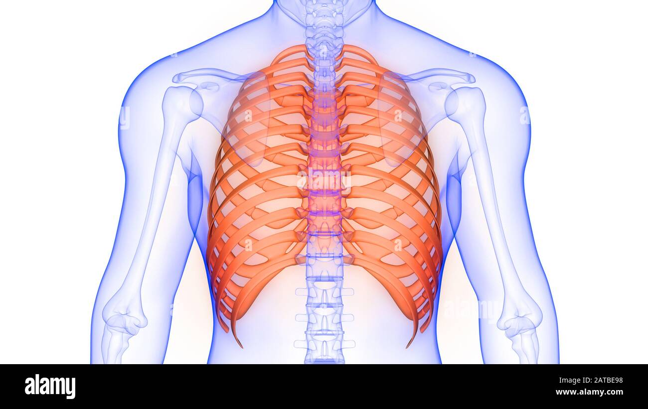
Vital organs such as heart and lungs are protected by the rib cage.
It is made up of 12 pairs of ribs. The heads of ribs 1, 10, 11, and 12 have a single facet for articulation with the bodies of the thoracic vertebrae. These muscles help the body bend at the waist. We think this is the most useful anatomy picture. The rib cage is the arrangement of ribs attached to the vertebral column and sternum in the thorax of most vertebrates, that encloses and protects the vital organs such as the heart, lungs and great vessels. It is also the center around which the superior 10 ribs directly or indirectly attached. The rib cage is often simplified as an oval shape. Don't be fooled their long, curved shape! Anatomynote.com found heart, lung, diaphragm and ribs location from plenty of anatomical pictures on the internet. Rib cage, in vertebrate anatomy, basketlike skeletal structure that forms the chest, or thorax, and is made up of the ribs and their corresponding attachments to the sternum (breastbone) and the vertebral column.the rib cage surrounds the lungs and the heart, serving as an important means of bony protection for these vital organs.in total, the rib cage consists of the 12 thoracic vertebrae and. On the trunk of the body in the thoracic area, the shoulder in general is the acromial, while the curve of the shoulder is the deltoid. Human anatomy for muscle, reproductive, and skeleton. Ribs 11 and 12 do not have necks or tubercles and the anterior tips of their bodies lack an articular surface.
Numbered ribs, sternum, cartilage parts and clavicular articulation.ribs eight to ten are the false ribs and are connected to the sternum indirectly via the cartilage of the rib above learn everything about the ribs with our articles, video tutorials, quizzes, and labeled diagrams there are eleven pairs of external intercostal muscles and these are. And more specifically, the rib cage is an egg with planes. The major muscles of the abdomen include the rectus. The muscles of the abdomen protect vital organs underneath and provide structure for the spine. We hope this picture heart, lung, diaphragm and ribs location can help you study and research.
Thoracic Spine from www.spineuniverse.com The primary responsibilities of the ribcage involve protecting the thoracic visceral organs, enclosing the thoracic visceral organs, and is included. The rib cage is more like an egg because the top is narrower than the bottom. On the trunk of the body in the thoracic area, the shoulder in general is the acromial, while the curve of the shoulder is the deltoid. For a gesture drawing, that's good enough. Our latest youtube film is ready to run. Vital organs such as heart and lungs are protected by the rib cage. The rib cage is often simplified as an oval shape. We hope this picture heart, lung, diaphragm and ribs location can help you study and research.
The intercostal nerves are part of the somatic nervous system, and arise from the anterior rami of the thoracic spinal nerves from t1 to t11.
Anatomy diagram rib area / pin on anatomy : Anatomy of ribs and its related area, find out more about anatomy of ribs and its related area. Its functions are to protect the thoracic organs from trauma and also form the bony attachment for various muscles. Anatomynote.com found heart, lung, diaphragm and ribs location from plenty of anatomical pictures on the internet. The intercostal nerves are part of the somatic nervous system, and arise from the anterior rami of the thoracic spinal nerves from t1 to t11. Rib 2 is thinner and longer than rib 1, and has two articular facets on the head as normal. On the trunk of the body in the thoracic area, the shoulder in general is the acromial, while the curve of the shoulder is the deltoid. Related posts of rib cage diagram with organs womens body parts stomach. It is made up of 12 pairs of ribs. The top edge of the manubrium has a depression called the suprasternal or jugular notch. Ribs 11 and 12 do not have necks or tubercles and the anterior tips of their bodies lack an articular surface. The bones of the rib cage are the sternum, the 12 thoracic vertebrae and the 12 pairs of ribs. The intercostal nerves are distributed chiefly to the thoracic pleura and abdominal peritoneum, and differ from the anterior rami of the other spinal nerves in that each pursues an independent course without plexus formation.
The rib cage is an important part of the human anatomy. We think this is the most useful anatomy picture. The cervicothoracic junction is where the neck (cervical spine) connects with the upper back (thoracic spine). The bones of the rib cage are the sternum, the 12 thoracic vertebrae and the 12 pairs of ribs. Thus, the sternal angle and second rib are important landmarks for the identification and counting of the lower ribs.
Rib Cage And Human High Resolution Stock Photography And Images Alamy from c8.alamy.com It is made up of 12 pairs of ribs. The right scapula from the front and back side. Since the first rib is hidden behind the clavicle, the second rib is the highest rib that can be identified by palpation. Just like in the manubrium, slight concave indentations in the lateral sides of the body of the sternum provide stronger attachment points for the costal cartilages to prevent rib separation. Anatomy of the rib cage diagram anatomy of the rib cage diagram in this image, you will find thoracic vertebrum, costochondral joint, costal cartilage, costal margin, costal arch, thoracic vertebrum, xiphoid process, xiphisternal joint, body, manubrial sternal joint, manubrium, the sternal notch in it. This bony framework plays an essential role in protecting the organs that lie in the thoracic region. The rib cage is more like an egg because the top is narrower than the bottom. For more anatomy content please follow us and visit our website:
Anatomy diagram rib area / pin on anatomy :
The head only articulates with the body of the t1 vertebra and therefore only one articulatory surface is present. Rib cage anatomy the rib cage, shaped in a mild cone shape and more flexible than most bone sets, is made up of varying elements such as the thoracic vertebra, 12 equally paired ribs, costal cartilage, and held together anteriorly by the sternum. Each pair is numbered based on their attachment to the sternum, a bony process at the front of the rib cage which serves as an anchor point. Rib bones are not classified as long bones.instead, anatomists classify the ribs as flat bones, and they are located within the axial skeleton.together with the sternum, thoracic vertebrae, and costal cartilages, the ribs form the thoracic cage, also called the bony thorax. The heads of ribs 1, 10, 11, and 12 have a single facet for articulation with the bodies of the thoracic vertebrae. Its functions are to protect the thoracic organs from trauma and also form the bony attachment for various muscles. Anatomynote.com found heart, lung, diaphragm and ribs location from plenty of anatomical pictures on the internet. The cartilage that forms at the end of each rib (costal cartilage) attaches either. Since the first rib is hidden behind the clavicle, the second rib is the highest rib that can be identified by palpation. The anatomy of the human ribs is made up of 24 ribs which are parted in 12 pairs (each on the left and right side of the chest wall), with the sternum, metasternum (the xiphoid process), and the costal cartilages all situated at the anterior of the chest wall, followed by the thoracic vertebrae on the posterior of the chest wall. Rib 1 is also flattened horizontally. The sternum is a flat bone that is made up of three parts, the (1) manubrium, (2) body, and the (3) xiphoid process. And more specifically, the rib cage is an egg with planes.


0 Comments:
Posting Komentar